IMDEA and UPM 3D print braided nitinol stents in a single step, hit 50% compression with 90% recovery
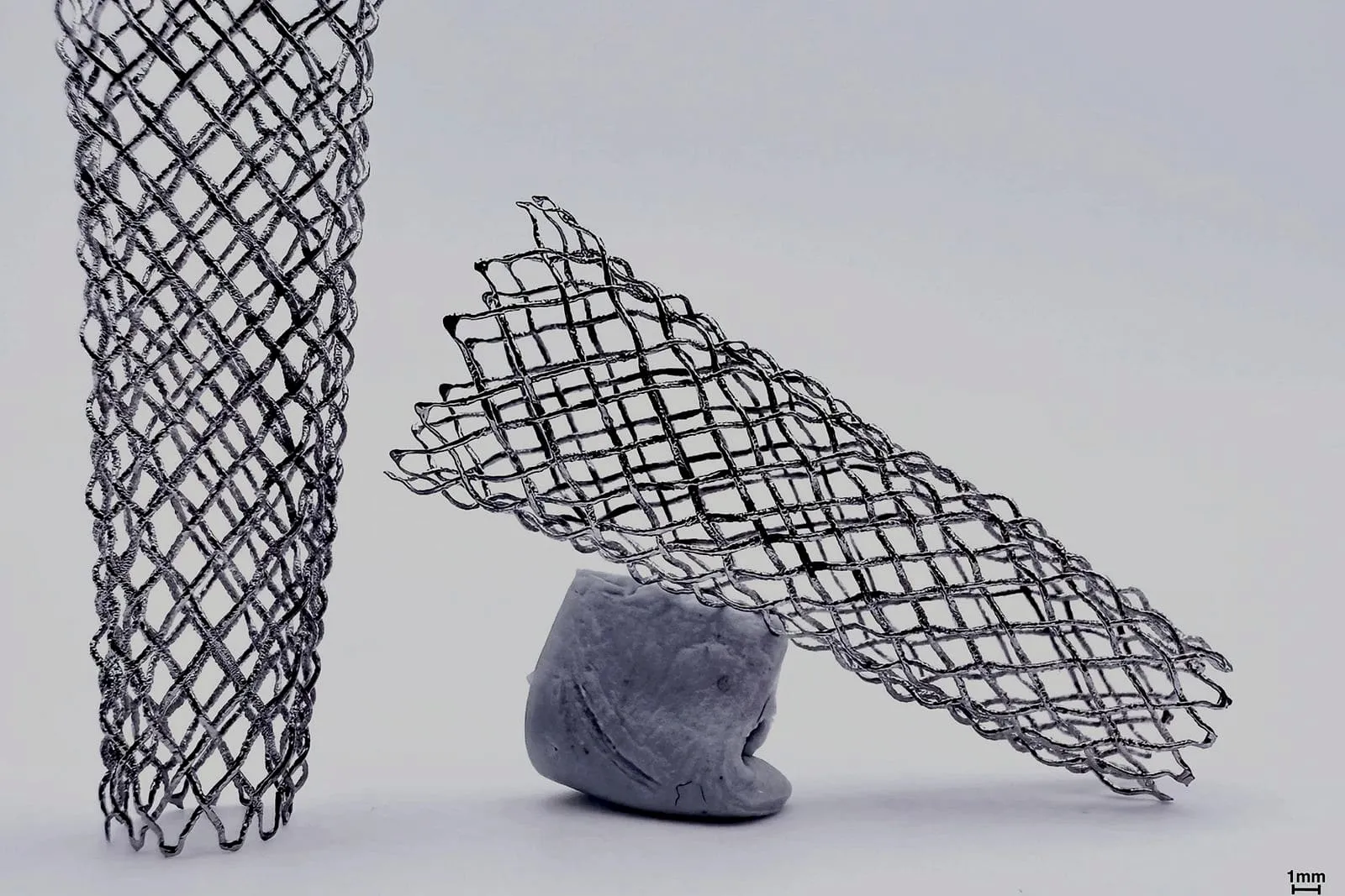
Researchers at the IMDEA Materials Institute and Universidad Politécnica de Madrid, with MIT and the University of Galway, have 3D printed braided nitinol structures for transcatheter devices in a single production step rather than weaving wire after the fact. The process has been patented and won the National James Dyson Award.
Most printed metal medical devices use lattices, and the team says that’s the wrong geometry for a stent that has to collapse into a catheter and spring back open. Lattices deform by bending rigid struts: compressed, they get stiff fast, push harder on the artery wall than intended, and the authors cite fracture risk approaching 25% strain. Braids deform instead by letting filaments slide and reorient against each other, which is closer to how woven nitinol stents already behave.
| Property | Measured value |
|---|---|
| Fiber thickness reduction (etching + electropolishing) | 47.7% |
| Cross-sectional area reduction | 71.1% |
| Maximum compression tested | 50% of original diameter |
| Elastic recovery after compression | >90% |
| Chronic outward force vs. conventional lattices | up to 5x lower |
Surface finish mattered enough that as-printed fibers needed chemical etching and electropolishing before the structures were usable; the numbers above are the before-and-after on that step. Validation so far is a single compression-expansion cycle per prototype, simulating catheter loading and release, which is a long way from a clinical device. The authors list in vitro biocompatibility and animal studies as the next required steps before any patient trial, and they flag neurovascular applications in hard-to-reach brain regions as a further target, which would need more miniaturization via two-photon polymerization with metallic suspensions and magnetic functionalization for remote navigation.