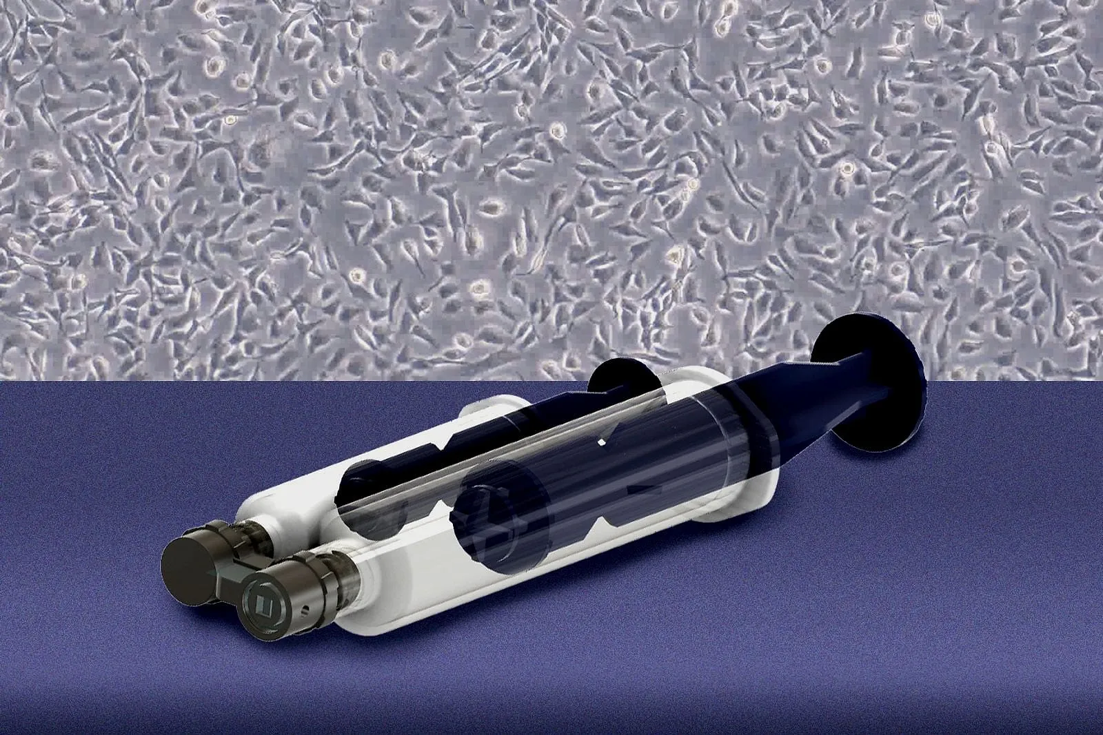
Researchers at MIT and Johns Hopkins University built a handheld device with a 3D printed microfluidic channel that pulls living cells off targeted spots on excised tissue, described in the journal Device and tested on fresh human fallopian-tube samples supplied by Johns Hopkins.
The problem it targets: high-grade serous ovarian cancer, the most common form of ovarian cancer, often starts in the fallopian tubes with precursor lesions too small to spot reliably. Standard pathology fixes removed tissue in preservative and slices it for viewing, which kills the cells and rules out culturing them. MIT professor Kripa Varanasi, who led the work, said the goal was to “collect them alive and study how they behave,” not just look at them under a microscope.
The device uses two syringes. One creates a vacuum that holds the 3D printed unit flush against the tissue and forms a seal so nothing leaks. The second pushes fluid through the microfluidic channel, confining flow to a small region of tissue so the shear stress from that flow detaches cells without destroying them. Postdoctoral researcher Bert Vandereydt, co-first author on the study, described it as a microfluidic chip that “gently shears the cells off.”
Against unspecified conventional collection methods, the team reports that cells pulled with the device stayed viable and grew in culture more readily. Using the fallopian-tube samples, they grew organoids from the collected cells, then shipped the remaining tissue back for standard pathology.
The researchers plan to keep working with tissue already removed from the body, which they say is the faster path to regulatory clearance, before attempting to use the device to swab cells inside a patient.
The source does not publish viability percentages, organoid growth rates, sample counts, channel dimensions, print material, or a timeline for in-patient testing.